In the complex world of cellular biology, calcium ions act as one of the most versatile and ubiquitous secondary messengers. They are responsible for a staggering array of biological processes, from the initial spark of fertilisation to the contraction of muscle fibres and the firing of neurons in the brain. Because calcium is so central to how cells communicate and respond to their environment, being able to observe its movement in real time is invaluable for researchers. This is where the calcium flux assay comes into play, providing a window into the dynamic internal life of a cell.
At its core, this assay measures the movement of calcium ions across the cell membrane or their release from internal stores like the endoplasmic reticulum. When a cell is stimulated—perhaps by a hormone, a neurotransmitter, or a potential drug candidate—calcium levels within the cytoplasm can spike rapidly. By monitoring these spikes, scientists can determine exactly how a cell is reacting to a specific stimulus. This isn’t just about whether a reaction happens; it is about the speed, intensity, and duration of that reaction, all of which provide critical clues about the underlying biological mechanisms.
How the calcium flux assay works in practice
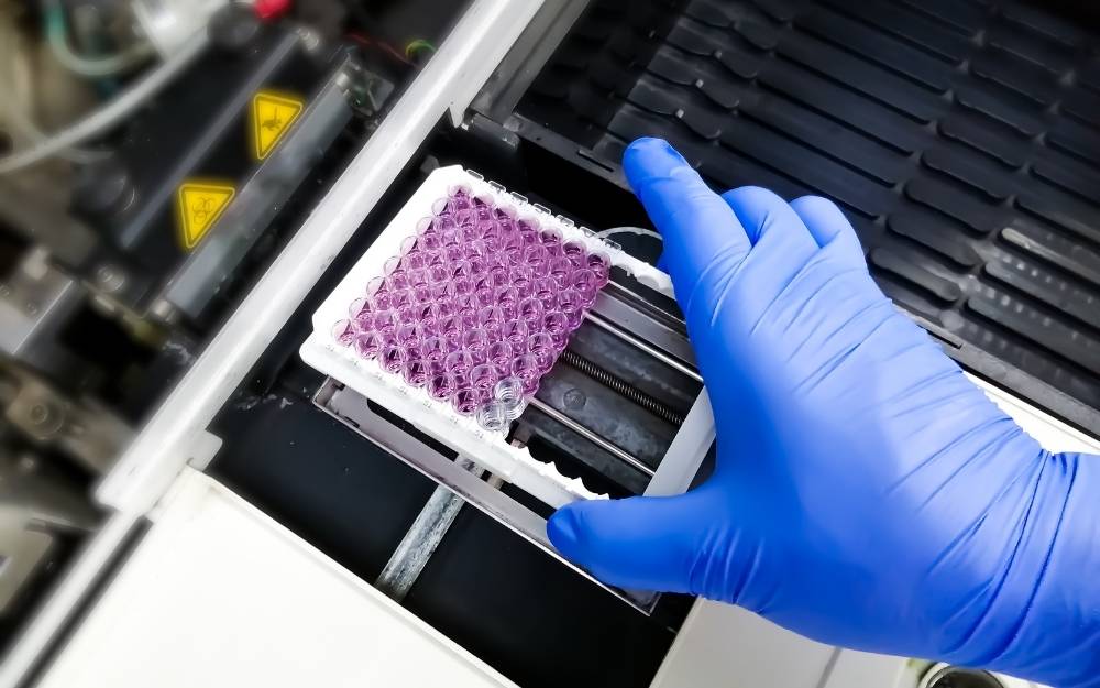
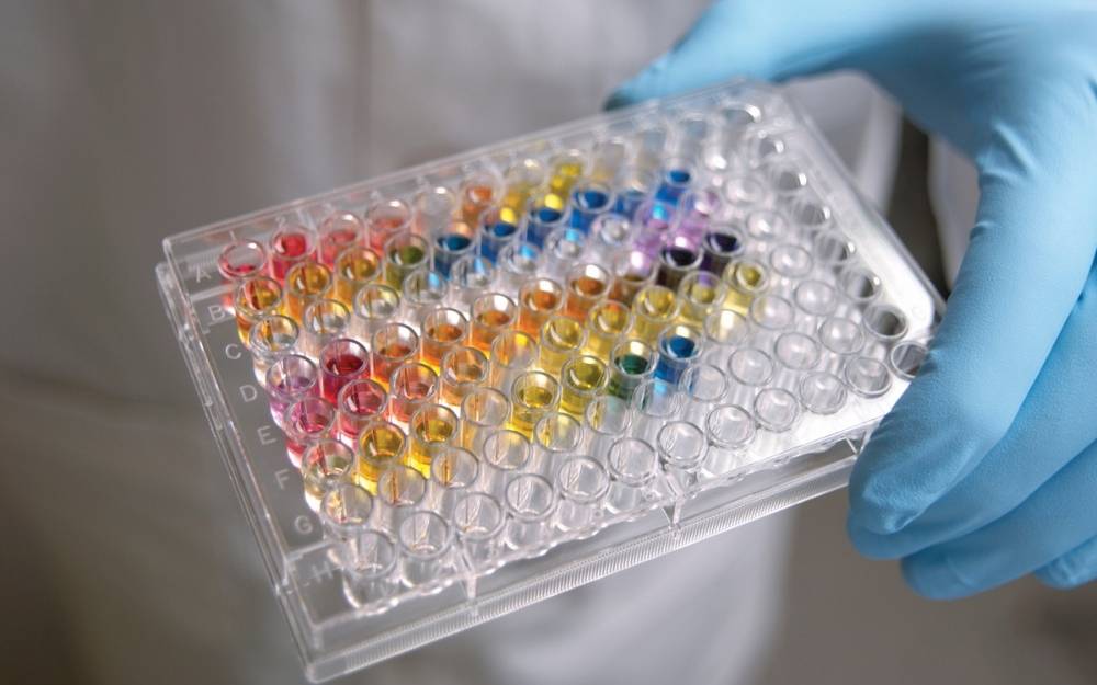
The beauty of a modern calcium flux assay lies in its sophisticated use of fluorescent indicators. These are specialised dyes or genetically encoded proteins that change their light-emitting properties when they bind to calcium ions. The process typically begins by loading these indicators into the cells of interest. Once the cells are ‘labelled’, they are placed into a controlled environment, often a multi-well plate, and exposed to various compounds.
As the calcium concentration increases, the fluorescence intensity changes, which is then captured by highly sensitive optical equipment. This data is recorded as a kinetic profile, showing a baseline of activity followed by a sharp peak and a gradual return to equilibrium. This real-time monitoring is what sets the assay apart from endpoint measurements, which only give a ‘before and after’ snapshot. With flux assays, you see the entire journey of the cellular response.
The importance of fluorescent indicators
Choosing the right indicator is a crucial step in ensuring the experiment is properly optimised. There are two main categories of dyes used in these assays:
- Single-wavelength indicators: Dyes like Fluo-4 or Fluo-8 are incredibly popular because they offer a very high increase in fluorescence intensity upon binding to calcium. They are easy to use and work well with standard plate readers, making them ideal for high-throughput screening.
- Ratiometric indicators: Dyes such as Fura-2 or Indo-1 are slightly more complex. They involve measuring fluorescence at two different wavelengths and calculating the ratio between them. This method is often preferred when researchers need to account for variations in dye loading, cell thickness, or photobleaching, as the ratio remains stable even if the absolute signal fluctuates.
Why researchers still rely on this method today
Despite the emergence of many new technologies in the lab, the calcium flux assay remains a staple in both academic research and the pharmaceutical industry. One of the primary reasons for its longevity is its incredible sensitivity. Because calcium signals are often the first thing to happen after a receptor is activated, the assay can detect responses that other methods might miss. It is also remarkably fast; many calcium responses occur within seconds, allowing for rapid data collection.
Furthermore, the assay is highly scalable. In the world of drug discovery, where thousands of compounds need to be tested against a single target, the ability to automate the process is vital. Modern robotic systems can run these assays in 384-well or even 1536-well formats, processing vast libraries of chemicals in a fraction of the time it would take using older techniques. This efficiency is a major driver in reducing the time and cost associated with bringing new medicines to market.
Practical applications in modern medicine
The versatility of the calcium flux assay means it finds a home in many different areas of research. Perhaps its most well-known application is in the study of G protein-coupled receptors (GPCRs). GPCRs are a massive family of proteins that sit on the cell surface and are the targets for roughly one-third of all FDA-approved drugs. When a drug binds to a GPCR, it often triggers a calcium release, making the flux assay the perfect tool for identifying new agonists or antagonists.
Beyond GPCRs, the assay is essential for several other key areas:
- Ion Channel Research: Scientists use these assays to study how ions move through specific channels in the cell membrane, which is critical for understanding heart rhythm, nerve function, and muscle health.
- Safety Pharmacology: Before a drug can move to human trials, it must be screened for potential toxicity. Monitoring calcium flux in cardiac cells (cardiomyocytes) can reveal if a drug might cause dangerous arrhythmias or other heart-related side effects.
- Neuroscience: By observing calcium oscillations in neurons, researchers can better understand how brain cells communicate and how diseases like Alzheimer’s or Parkinson’s disrupt these signalling pathways.
Navigating the technical hurdles of the assay
While the concept is straightforward, executing a perfect calcium flux assay requires careful attention to detail. There are several factors that can interfere with the results if not properly managed. For instance, ‘background noise’ from the medium or the compounds themselves can sometimes mask the fluorescent signal. To combat this, many laboratories utilise ‘no-wash’ assay kits that include quenching dyes to suppress unwanted background fluorescence.
Another challenge is the behaviour of the cells themselves. Cells must be healthy, at the right stage of growth, and handled gently to ensure they respond naturally to the stimuli. Temperature control is also vital; since biological processes are temperature-dependent, even a small fluctuation can change the kinetics of the calcium response and lead to inconsistent data. Ensuring that the equipment is calibrated and the environment is stable is a full-time job for many lab technicians.

Optimising the protocol for better data
To get the most out of a calcium flux assay, it is often necessary to refine the protocol for each specific cell type or target. This might involve adjusting the concentration of the dye, the incubation time, or the buffer solution used during the experiment. Many researchers also find that using specialised plate coatings can help the cells adhere better, which is particularly important when using automated liquid handling systems that might otherwise dislodge the delicate cell layer.
By paying close attention to these variables, scientists can produce high-quality, reproducible data that provides a clear picture of cellular activity. This level of precision is exactly what is needed when making the high-stakes decisions involved in drug development, where a single ‘false positive’ or ‘false negative’ result can have significant financial and scientific consequences.
Interpreting the data for better results
Once the assay is complete, the focus shifts to data analysis. Modern software can automatically calculate several different parameters from the kinetic curves, such as the peak height (Emax), the time it takes to reach that peak, and the area under the curve (AUC). Each of these metrics tells a different part of the story. For example, a drug that produces a very high peak might be a potent activator, while a drug that produces a lower, sustained signal might be acting through a different, more subtle mechanism.
Comparing these results across different concentrations of a drug allows researchers to create dose-response curves. These curves are the ‘gold standard’ for determining the potency and efficacy of a potential new treatment. By analysing how the calcium flux changes as the dose increases, scientists can identify the ‘sweet spot’ where a drug is most effective without being toxic. This quantitative approach is what transforms a simple observation of light into a powerful tool for medical progress.

Cinthia is a freelance journalist with a passion for current affairs, lifestyle trends, and expert advice. She delivers well-researched news and actionable tips to keep readers informed and engaged.